Medical & Surgical Vitreoretinal Services
Retina is the light receptive screen of the eye (akin to the screen in the camera) that sends visual messages to the brain through the optic nerve. The vitreous is a gel like substance filling the eye behind the lens and in front of the retina. It is an optically clear viscous medium. The vitreous can be become opaque due to bleeding in the eye due to injury, diabetes mellitus etc.
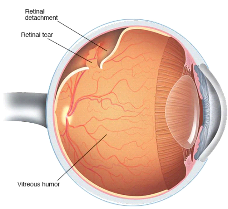
Retinal Detachment
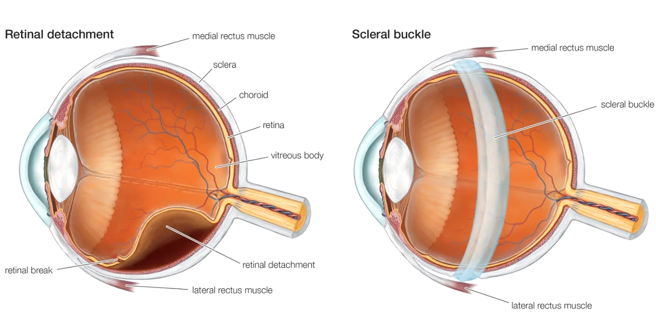
When the retina is lifted or pulled from its normal position it gets detached. This interferes with the reception of light by the retina. If this is not treated immediately, this could result in permanent blindness. Retinal detachments are caused by a host of conditions in the eye.
Rhegmatogenous detachment – caused by retinal tears or breaks (dehiscence in the retina). Rhegmatogenous detachment is the most common type.
Tractional detachment caused by tractional bands (due to fibrous tissue growth on the surface of retina).
Exudative detachment caused by injury to or inflammation in the eye (when the retina is lifted by fluid accumulation behind it)
Retinal detachment can occur if you
Are over 40 years of age
Are myopic (especially high myopia)
Had a retinal detachment in one eye
Have a family history of retinal detachment
Have other eye diseases like retinoschisis, Uveitis, lattice degeneration, retinal breaks etc
Had an eye injury
Untreated vitreous haemorrhage, diabetic retinopathy etc.
Symptoms
Sudden appearance or increase in “Floaters“ – black cobweb like little specks in the field of vision
Flashes of light
Appearance of a curtain over the field of vision
Sudden or gradual drop in vision
Diagnosis of retina and vitreous diseases is done with.
Indirect Ophthalmoscopy
B – Scan ultrasound
Optical Coherence Tomography
Digital Fundus camera
Treatment
Laser indirect Ophthalmoscopy – for retina breaks in which the hole or break in the retina is sealed with laser
Cryopexy – which freezes the area around the hole to reattach the retina
Pnuemoretinopexy – where gas is injected into the vitreous to push the retina to its normal position
Scleral buckling – where a buckle is attached to the outside of the eye to gently push the wall of the eye against the detached retina
Vitrectomy – is performed with the state of the art Vitrectomy machine in which the vitreous is removed partly or totally through a small incision in the sclera, gas is injected into the eye to push the retina to its normal position
Over 90% of retinal detachment can be successfully treated with the good visual outcomes achieved when the patient presents early in the course of the disease. It is vital to report to the eye specialist at the first instance of noticing floaters, flashes or curtains in the field of vision.
AMARDEEP EYE CARE is fully equipped for the management of disorders of the retina and vitreous with very experienced vitreoretinal surgeons providing all the available treatment options. AMARDEEP EYE CARE also has a – state – of – the – art machines and Operation theatres dedicated to treating vitreoretinal conditions.
Diabetic Retinopathy
Diabetes affects almost 40% of the general population and can affect every part of the body. Similarly it can affect every part of the eye. Individuals with uncontrolled blood sugar can have frequent allergies of the eye and boils in the eyelids. Diabetes can also cause cataract to occur at an earlier age than normal.
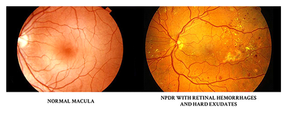
The nerve layer of the eye called the retina functions like the film of the camera. The serious eye defect due to diabetes occurs in the retina. This is called diabetic retinopathy. Individuals at high risk for Diabetic Retinopathy are those who have poorly controlled blood sugar values for a long time, kidney disorders, blood pressure, anaemia and high cholesterol levels.
The initial stages of the disease are usually asymptomatic where the presence of Diabetic Retinopathy can only be picked up by an Ophthalmologist after a thorough examination of the eyes. It is only when the disease is very advanced that vision is affected permanently. Initial symptoms, when present, include poor vision and difficulties in reading. Advanced disease results in complete permanent loss of vision.
Every individual with diabetes must have an eye check-up done at least once in a year. Early identification of disease and treatment with lasers or injections are capable of preserving vision but advanced disease results in permanent vision loss. This loss of vision in diabetic retinopathy can be easily prevented if the disease is diagnosed in its early stages.
AMARDEEP EYE CARE is equipped with the most advanced screening devices which are used to identify diabetic retinopathy such as Optical coherence tomography (OCT), Fundus Fluorescein angiography (FFA) and Fundus photography.
At AMARDEEP EYE CARE state-of-the-art treatment options with retinal laser therapy, injections in the eye (Ranibizumab, Aflibercept) and implants in the eye (Ozurdex) are done as an out-patient procedure. Advanced retinopathy is also managed with the Constellation surgical system and the Leica ophthalmic surgical microscope. With experienced professionals and state of the art technology, treatment is carried out with ease and precision to deliver excellent and promising results.